

ECG Machines and Accessories
An ECG (electrocardiogram) machine is a non-invasive device used to measure and record the electrical activity of the heart. Williams Medical Supplies offers ECG machines and accessories for UK primary care.
More Information
ECG Machines
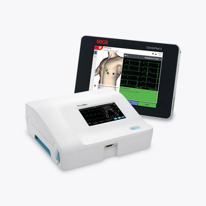
Williams Supplies offers a range of ECG machines. From desktop and PC based with built in printers, handheld ECGs, accessories, and paper to Holter machines for long term monitoring.

Accessories
Our range of trolleys, cases, paper and electrodes to pair with your ECG Machine
How an ECG Machine Works An ECG machine measures the electrical activity of the heart through electrodes placed on the skin. Each heartbeat is triggered by an electrical impulse that travels through the cardiac conduction system. The ECG machine captures and records these impulses as a waveform, displaying a characteristic pattern for each heartbeat. A standard ECG trace contains three main components. The P-wave represents electrical activity in the atria as they contract to push blood into the ventricles. The QRS complex represents electrical activity in the ventricles as they contract to pump blood to the lungs and body. The T-wave represents ventricular recovery, when the ventricles reset electrically ready for the next heartbeat. The ECG machine records these signals through a patient cable connected to adhesive electrodes placed on the chest, arms, and legs. A 12-lead ECG machine uses 10 electrodes to generate 12 views of cardiac electrical activity from different angles. Each view is called a lead. Together, the 12 leads give a clinician a detailed map of how electrical signals move through the heart. Types of ECG Machine ECG machines are available in several configurations, each suited to different clinical environments and diagnostic requirements. 12-Lead Desktop ECG Machines A 12-lead desktop ECG machine is the standard device for resting ECG recording in primary care, hospitals, and community clinics. It records 12 simultaneous views of cardiac electrical activity using 10 electrodes placed on the chest, wrists, and ankles. Desktop machines typically feature a built-in display, thermal or laser printer, and clinical system integration via USB or network connection. Most UK general practice ECG machines support connectivity with EMIS, SystmOne, and Vision through compatible software. Williams Medical Supplies supplies desktop ECG machines from leading clinical brands, with options available for single-room and multi-room practice use. Portable and Holter ECG Monitors Portable ECG monitors allow continuous cardiac monitoring over 24 to 48 hours or longer. A Holter monitor is worn by the patient and records every heartbeat across the monitoring period, capturing arrhythmias that may not be present during a short resting ECG. Portable monitors are used for palpitation investigation, syncope assessment, and monitoring patients with known arrhythmias on medication. Results are downloaded and analysed by a clinician after the monitoring period. Personal and Single-Lead ECG Devices Personal ECG devices, such as the AliveCor KardiaMobile, record a single-lead ECG in 30 seconds using a smartphone app. They are used in primary care for opportunistic atrial fibrillation screening and by patients for remote and between-appointment monitoring. The KardiaMobile is FDA-cleared and CE-marked and is referenced in NICE guidance on AFib detection. Personal ECG devices require no gels, wires, or clinical preparation. What Can an ECG Detect? An ECG machine records the electrical activity of the heart at the time of the recording. It does not measure blood flow, valve function, or structural anatomy directly. Within those parameters, an ECG can identify a range of cardiac conditions and abnormalities. Common conditions identifiable from an ECG recording include: Arrhythmias: abnormal heart rhythms including atrial fibrillation, atrial flutter, supraventricular tachycardia, ventricular tachycardia, and heart block. Conduction abnormalities: including left bundle branch block (LBBB), right bundle branch block (RBBB), and first, second, and third degree heart block. Signs of myocardial infarction: ST elevation, ST depression, pathological Q-waves, and T-wave inversion may indicate acute or previous myocardial infarction. Left ventricular hypertrophy (LVH): increased QRS voltage and axis deviation associated with longstanding hypertension or aortic stenosis. Electrolyte disturbances: hyperkalaemia, hypokalaemia, and hypercalcaemia each produce characteristic ECG changes. Medication effects: QT prolongation and other changes associated with antiarrhythmic drugs, antipsychotics, and other QT-affecting medicines. An ECG does not detect coronary artery disease in the absence of ischaemia, heart failure, structural abnormalities, or intermittent arrhythmias that are not present at the time of recording. Additional investigations such as echocardiography, exercise ECG, or ambulatory (Holter) monitoring may be required where the clinical picture is not resolved by a resting 12-lead ECG. When is an ECG Used in Primary Care? ECG recording is a core diagnostic function in UK general practice. Common indications for ECG in primary care include: Chest pain assessment: to identify ischaemic changes, arrhythmia, or conduction abnormality in patients presenting with chest pain or discomfort. Palpitation investigation: to capture rhythm disturbances in patients reporting palpitations, racing heart, or irregular heartbeat. Pre-operative assessment: many surgical procedures require a baseline resting ECG, particularly in older patients or those with cardiovascular risk factors. Hypertension monitoring: to screen for left ventricular hypertrophy in patients with longstanding or uncontrolled high blood pressure. Medication monitoring: patients taking drugs with cardiac effects, including digoxin, amiodarone, antipsychotics, and certain antidepressants, may require regular ECG monitoring. Atrial fibrillation screening: NICE guidance recommends opportunistic ECG screening for AFib in patients aged 65 and over, and in patients with additional cardiovascular risk factors. Syncope and collapse investigation: to identify arrhythmias or conduction defects in patients presenting with unexplained collapse or loss of consciousness. Shortness of breath assessment: to support the differential diagnosis in patients with unexplained breathlessness or reduced exercise tolerance. Understanding ECG Results A normal resting ECG in an adult shows a regular sinus rhythm with a heart rate of 60 to 100 beats per minute. Each P-wave is followed by a QRS complex. The QRS duration is under 120 milliseconds. The PR interval is between 120 and 200 milliseconds. The T-waves are upright in most leads. Common ECG findings that may require clinical review or onward referral include: Atrial fibrillation: absent P-waves with an irregularly irregular ventricular response. This is the most common significant arrhythmia identified by ECG in primary care. Left bundle branch block (LBBB): QRS duration above 120ms with a broad, notched QRS in lateral leads. New LBBB in a patient with chest pain is treated as an acute presentation. ST elevation: may indicate acute myocardial infarction requiring immediate referral. Prolonged QT interval: QTc above 440ms in men or 460ms in women may indicate medication toxicity, electrolyte disturbance, or inherited channelopathy. Left ventricular hypertrophy: increased QRS voltage with or without repolarisation changes, often associated with hypertension. ECG interpretation requires clinical training and should be performed in the context of the patient's symptoms, history, and other investigations. An ECG result labelled as normal does not exclude all cardiac pathology. ECG Machine Accuracy and Limitations ECG machines record electrical signals accurately when electrodes are correctly placed and good skin contact is maintained. Several factors can affect the quality and clinical reliability of an ECG recording. Factors that may affect ECG accuracy include: Electrode placement errors: incorrect positioning of chest or limb electrodes is a common cause of abnormal-appearing ECG traces. Misplacement of V1 and V2 electrodes can produce appearances that mimic right bundle branch block or anterior ischaemia. Patient movement and muscle artefact: voluntary or involuntary movement during recording introduces electrical noise into the trace, particularly in anxious, shivering, or tremulous patients. Electrical interference: nearby electrical equipment can introduce 50Hz mains interference into an ECG recording. Poor electrode contact: dry, oily, or very hairy skin reduces electrode contact and degrades signal quality. Good skin preparation improves recording quality. Body habitus: obesity, emphysema, and pericardial effusion can reduce ECG voltage and affect lead appearances. Intermittent arrhythmia: a resting ECG records only the rhythm present at the time of the recording. A normal resting ECG does not exclude intermittent arrhythmia. Automatic ECG interpretation software is available on many desktop machines and is useful as a screening aid. Automatic interpretation should always be reviewed by a trained clinician before acting on the result. ECG Machine Servicing, Calibration, and Care Plans ECG machines used in clinical settings require regular servicing and calibration to maintain diagnostic accuracy and comply with clinical governance requirements. Williams Medical Supplies offers test and calibration services for ECG machines across primary care and community clinical settings. Our Seca ECG Care Plan provides a structured maintenance and support package for Seca ECG equipment. Care plans cover planned preventive maintenance, calibration verification, and equipment support, helping practices meet CQC inspection requirements for medical device maintenance. Regular servicing reduces unexpected equipment downtime and extends the working life of the device. Practices purchasing ECG equipment through Williams Medical Supplies can request a quote for a care plan or calibration service by contacting our team on 01685 846666 for further details. Our ECG Range at Williams Medical Supplies Williams Medical Supplies offers a range of medical-grade ECG machines for UK primary care, community clinics, and hospital outpatient settings. Our range includes 12-lead desktop ECG machines, portable monitors, and personal single-lead devices including the AliveCor KardiaMobile. Personal ECG devices and Desktop 12-lead machines are available across a range of price points to suit single-room GP practices through to multi-room clinical environments. Our support team are available to assist with specifications, servicing requirements, and care plan options. We offer free delivery on orders over £100, and next-day delivery on qualifying orders, with a UK-based after-sales support. Frequently Asked Questions: ECG Machines Can anxiety affect an ECG? Several medical sources state that anxiety can influence ECG results by changing heart rate and rhythm and by causing artefact. GE HealthCare notes that test related nervousness or chronic anxiety may be associated with ECG abnormalities, including rhythm irregularities and T wave inversion, and can sometimes contribute to false positive abnormal readings. Baptist Health reports that anxiety disorders and temporary anxiety can potentially affect EKG results, although unusual sensations do not always translate into ECG changes. A review on psychological stress and ECG changes published on the US National Institutes of Health database reports that stress can affect ECG parameters and may promote arrhythmias in susceptible people. What is a normal ECG? The British Heart Foundation describes a normal ECG as a tracing where the heart rate is typically between 60 and 100 beats per minute at rest, with a regular rhythm and consistent P waves, QRS complexes, and T waves in each heartbeat. The National Health Service (NHS) explains that in a normal ECG, electrical activity follows a regular pattern that shows the atria and ventricles contracting in sequence and does not display abnormal intervals or segments such as prolonged QT or significant ST elevation or depression. The American Heart Association adds that a normal ECG shows standard intervals, including a normal PR interval and QRS duration, and no evidence of ischemia or arrhythmia. What is the most common abnormality of an ECG? Health Awareness describes atrial fibrillation (AF) as the most common sustained heart rhythm abnormality, affecting an estimated 1.4 million people in the UK, and notes that AF is typically diagnosed using an ECG that shows an irregularly irregular rhythm with absent P waves. The British Heart Foundation also lists atrial fibrillation as the most common arrhythmia, explaining that it is frequently identified on an ECG by its irregular rhythm and can increase the risk of stroke. A review in the journal Heart reports that AF is the most common sustained cardiac arrhythmia seen in clinical practice, accounting for a large proportion of abnormal ECG findings in adults. Do GP surgeries have ECG machines? The NHS explains that ECG tests are widely available in the UK and can be performed in hospitals, community clinics, and some GP surgeries, indicating that many but not all GP practices have ECG equipment. The British Heart Foundation also notes that ECGs are sometimes done at a GP surgery or local health centre before referral to a hospital specialist if needed. Guidance from the Royal College of General Practitioners on cardiovascular diagnostics reports that many UK general practices now have 12 lead ECG machines to support primary care assessment of chest pain and palpitations, though access can still differ between practices. Disclaimer: The information on this page is intended for qualified healthcare professionals and is provided for general clinical reference purposes only. It does not constitute medical advice and should not replace clinical judgement, locally agreed protocols, or national guidance. Williams Medical Supplies is a medical equipment supplier and does not provide clinical diagnoses or treatment recommendations. Always refer to current NICE guidance, local clinical pathways, and your professional governing body for clinical decision making.